Verhoeff Van Gieson Stain: A Powerful Tool for Histological Examination
The Verhoeff Van Gieson stain, also known as VVG, is a histological staining technique used to visualize elastic fibers in tissue samples. This staining method has been widely used in medical research and diagnostics to study the structure and composition of connective tissue. By highlighting elastic fibers, the Verhoeff Van Gieson stain provides valuable information about the organization and integrity of tissues, which is essential for understanding various diseases and developing effective treatments.
Context and History
The Verhoeff Van Gieson stain was first introduced by Joseph Verhoeff, an American pathologist, in the early 20th century. Since then, it has become a standard technique in histology laboratories around the world. The stain combines two dyes, Verhoeff's elastic stain and Van Gieson's stain, to selectively stain elastic fibers and collagen, respectively. This combination allows for a clear visualization of the relationship between elastic fibers and other tissue components.
Details of the Staining Technique
The Verhoeff Van Gieson staining technique involves a series of steps, including fixation, dehydration, and staining. The tissue sample is first fixed to preserve its structure, and then dehydrated to remove water. The sample is then stained with the Verhoeff's elastic stain, which binds to elastic fibers, followed by Van Gieson's stain, which stains collagen and other tissue components. The resulting stained sample can be examined under a microscope to visualize the elastic fibers and their relationship to other tissue structures. 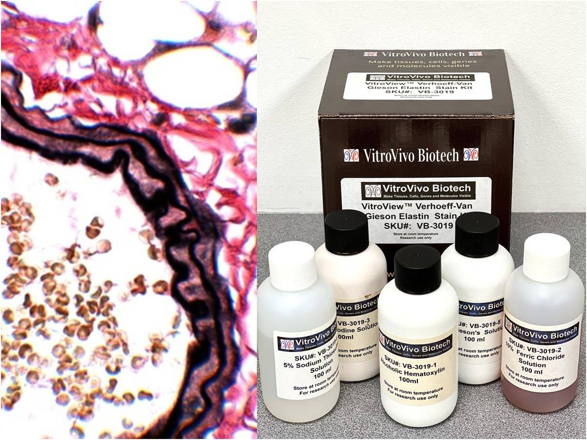
Implications and Applications
The Verhoeff Van Gieson stain has a wide range of applications in medical research and diagnostics. It is commonly used to study the pathology of cardiovascular diseases, such as atherosclerosis, where the integrity of elastic fibers plays a crucial role. The stain is also used to examine the structure of skin, lungs, and other organs, where elastic fibers are abundant. In addition, the Verhoeff Van Gieson stain is used in cancer research to study the relationship between tumor growth and the surrounding tissue structure. 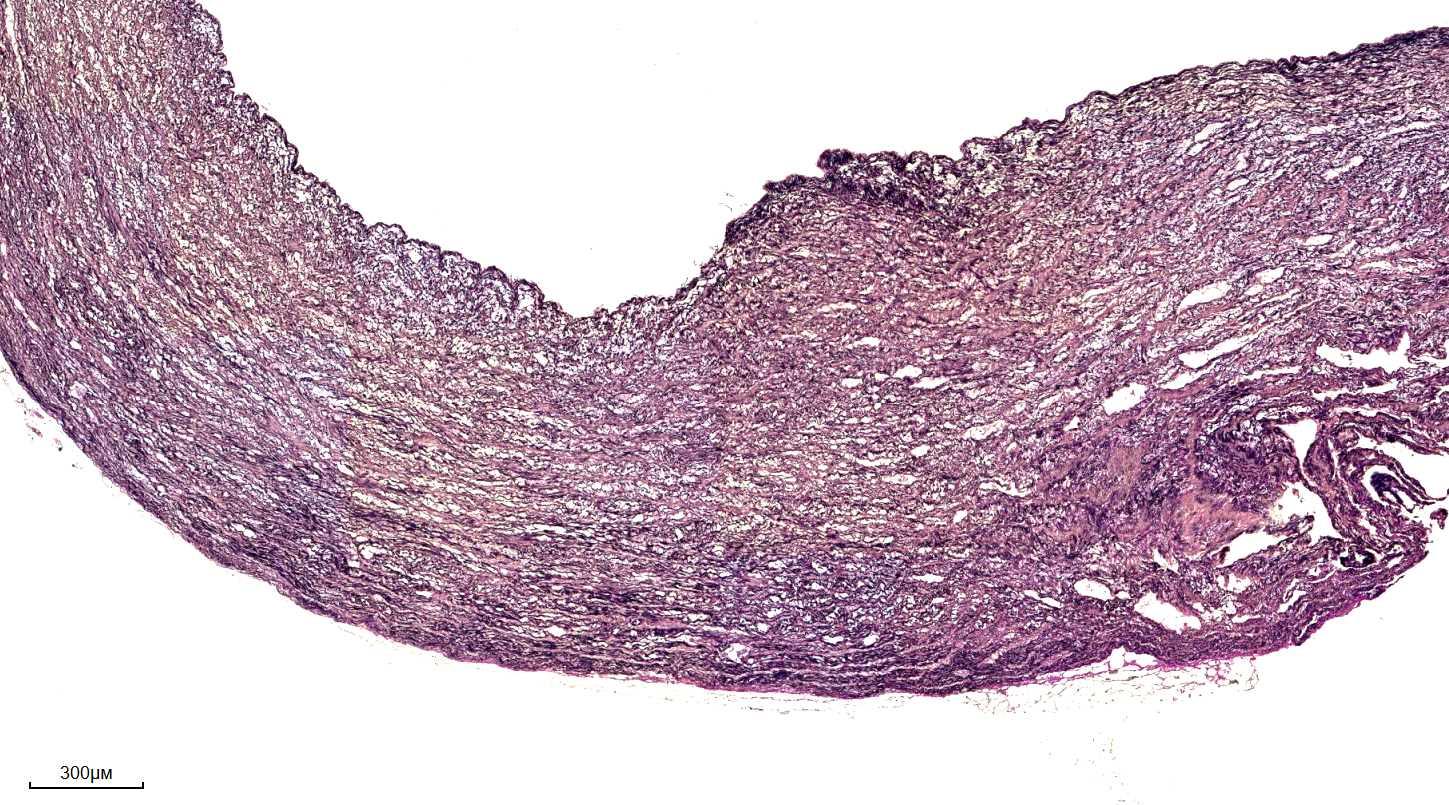
Benefits and Limitations
The Verhoeff Van Gieson stain offers several benefits, including its ability to selectively stain elastic fibers, which allows for a detailed examination of tissue structure. The stain is also relatively easy to perform and interpret, making it a valuable tool for researchers and clinicians. However, the stain also has some limitations, such as its sensitivity to fixation and staining conditions, which can affect the quality of the results. Additionally, the stain may not be suitable for all types of tissue samples, and alternative staining techniques may be required in some cases.
Future Directions
Despite its widespread use, the Verhoeff Van Gieson stain is still an actively researched topic, with ongoing efforts to improve its sensitivity and specificity. New staining techniques and imaging methods, such as fluorescence microscopy, are being developed to enhance the visualization of elastic fibers and other tissue components. These advances are expected to further expand the applications of the Verhoeff Van Gieson stain and provide new insights into the structure and function of tissues.
VitroView™ Verhoeff Van Gieson Elastin Stain Kit
VitroView™ Verhoeff Van Gieson Elastin Stain Kit
Verhoeff-van Gieson Elastic Stain Flashcards | Quizlet

Verhoeff-van Gieson Elastic Stain Flashcards | Quizlet
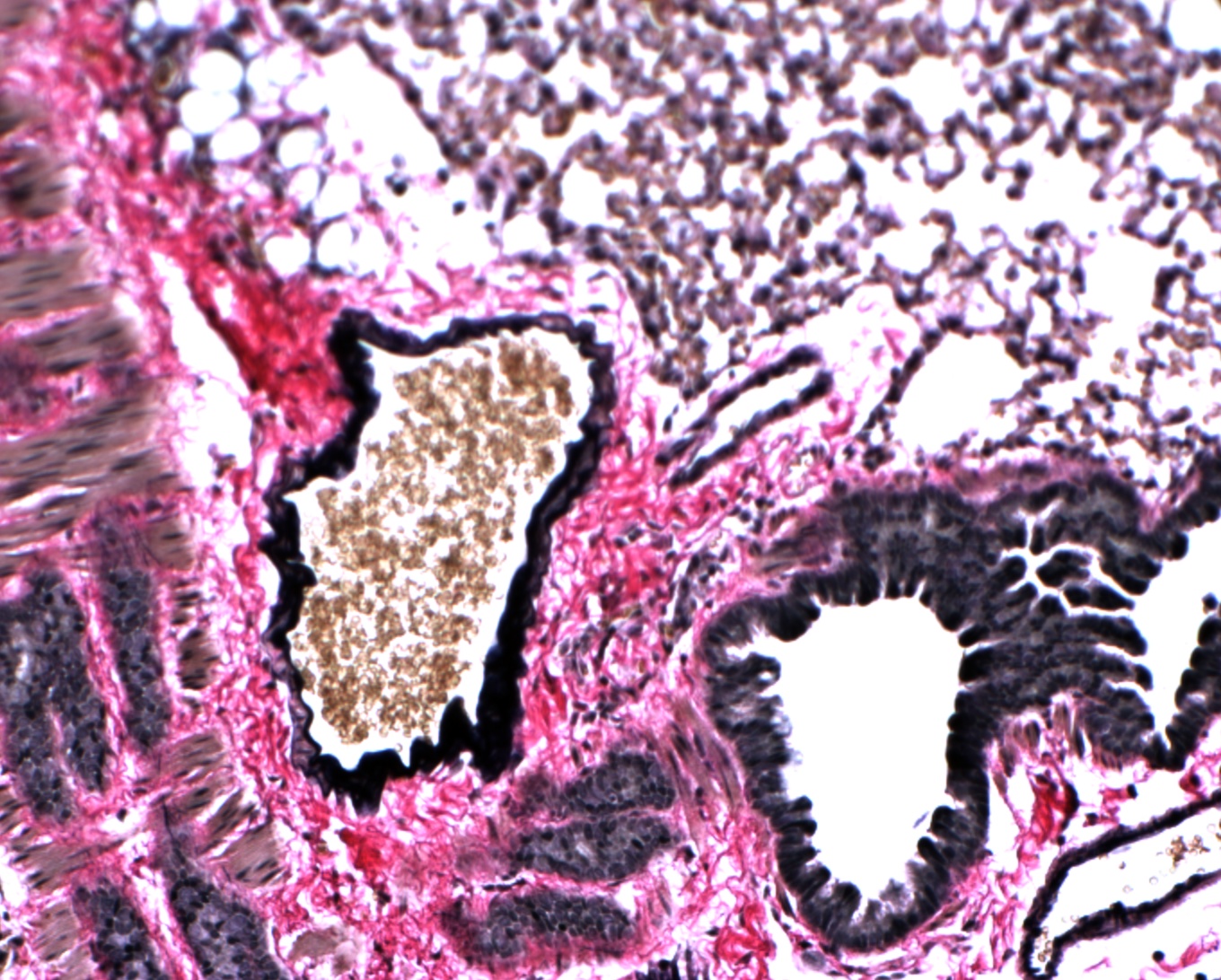
VitroView™ Verhoeff Van Gieson Elastin Stain Kit
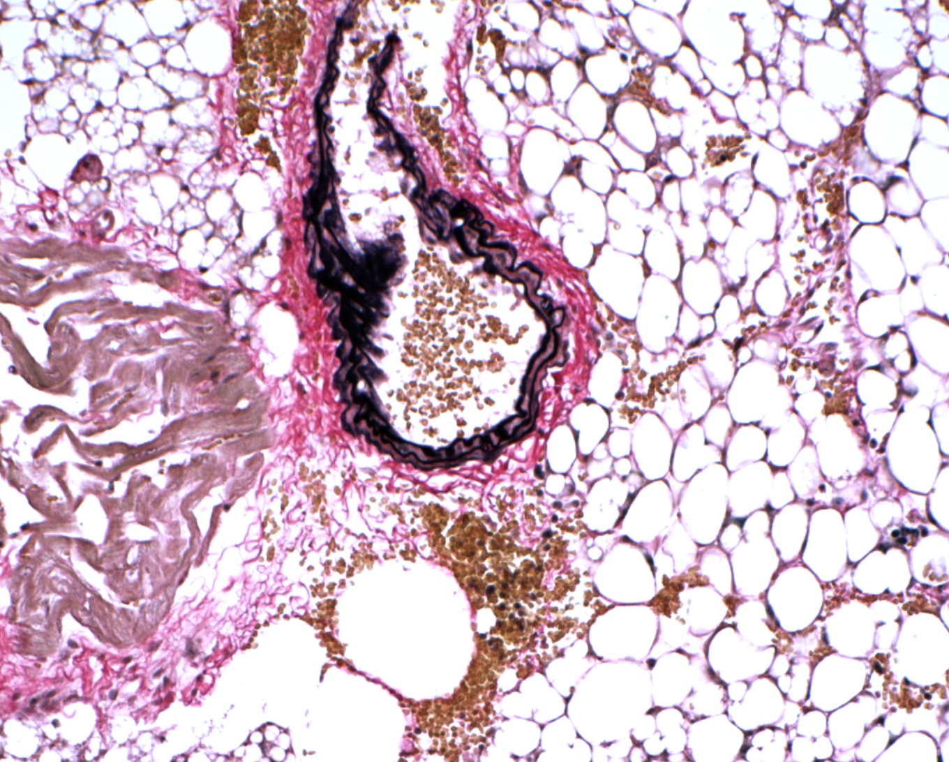
VitroView™ Verhoeff Van Gieson Elastin Stain Kit